Preserving “Peste des petits ruminants” virus with Whatman paper
- Post by: wsmib-admin
- mai 30, 2024
- Comments off
Preserving “Peste des petits ruminants” virus with Whatman paper
Akakpo Oluwole Christophert1*, Bodjo Sanne Charles2, Diallo Safiatou, Lamine3
1Department of Veterinary Vaccine Production and Quality Control, Faculty of Veterinary Medicine,
Pan African University life and Earth Science Institute (including Health and Agriculture), University of
Ibadan, Ibadan, Nigeria
2African-Union Pan African Veterinary Vaccine Center (AU-PANVAC), Bishoftu, Ethiopia
3Department of Veterinary Medicine, Institut Supérieur des Sciences et de Médecine Vétérinaire de
Dalaba (ISSMV), Dalaba, Guinée Conakry
Abstract
Introduction
“Peste des petits ruminants” virus (PPRV) poses a significant threat to livestock in Africa, where hot climates and limited cold chain infrastructure can hamper sample collection and transportation for diagnosis. This study evaluated the potential of Whatman Grade 3MM CHR chromatography paper as a tool for preserving PPRV nucleic acid at various temperatures.
Methods
PPR vaccine was serially diluted and spotted onto Whatman paper pieces. The paper samples were then stored at -20°C, +4°C, +24°C, +37°C, and +45°C for 31 days. RNA extraction and RT-PCR were performed to assess PPRV nucleic acid preservation.
Results
We found that Whatman paper effectively preserved undiluted (100) PPRV nucleic acid even at +45°C for the entire incubation period. However, for the 10-1 dilution, successful preservation was observed only at -20°C and +4°C. Under +24°C, +37°C, and +45°C, the 10-1 dilution remained stable for only the initial three days.
Conclusion
Whatman paper demonstrated promise as a method for transporting PPRV samples, particularly in resource-limited regions with high ambient temperatures. The paper can effectively preserve PPRV nucleic acid. Further research is needed to quantify the PPRV storage capacity of Whatman paper, allowing for optimization of sample volumes for transport and preservation purposes.
Keywords
Whatman paper, “Peste des petits ruminants” virus, Nucleic acid, RT-PCR
Introduction
“Peste des petits ruminants” (PPR) is a viral disease that primarily affects goats, sheep, camels and certain wild relatives of domesticated small ruminants. It is caused by a Morbillivirus closely related to the rinderpest virus. PPR was first reported in 1942 in Côte d’Ivoire (1).
The disease causes widespread illness and death in small ruminant populations, especially in regions of Africa, the Middle East, and Asia where these animals are crucial for people’s well-being. Sick animals exhibit high fever, depression, eye and nose discharges. Animals are unable to eat because their mouths become covered in painful erosive lesions, and they suffer from severe pneumonia and diarrhea. Death is a common outcome (2). PPR is classified into three pathogenic forms: per-acute, acute, and sub-acute.
However, the acute form is the most common, accounting for 80-90% mortality in individual flocks. When
the host is infected with Peste des petits ruminants virus (PPRV), general viraemia develops within 4-6 days, followed by high fever (40-41°C), marked salivation, shallow erosions in the oral mucosa, serous to purulent oculo-nasal discharge, dyspnea, coughing, pneumonia, and diarrhea. In the later stages, a sub-normal temperature of 37-38°C, combined with dehydration from diarrhea, can result in hypovolemic shock and the death of the affected animals (3).
PPR is diagnosed in the field primarily based on clinical signs, symptoms, and postmortem findings. Laboratory techniques are used to confirm the PPR diagnosis. Swabs from nasal secretions, mouth and rectal lining, conjunctival sac, clotted blood, and whole blood are usually collected from sick animals (with EDTA anticoagulant). Tonsils, tongues, spleens, lymph nodes, and affected areas of the alimentary tract mucosa may be collected post-mortem. Clinically PPR should be differentiated from other diseases
such as foot and mouth disease, contagious caprine pleuropneumonia, bluetongue, sheep and goat pox, and contagious ecthyma, which have similar symptoms (4). As a result, laboratory techniques provide an accurate and definitive diagnosis. PPR in clinical and pathological specimens can be diagnosed using a variety of techniques. These techniques are divided into two categories based on the type of substance detected and quantified: viral antigen and genome (whole virus, viral protein, or genome), and specific antibodies in serum (5). Filter paper has been used more widely as a method for sample collection in field
specimens (6).
Filter papers have been shown to be suitable for the long term preservation of either DNA or RNA viruses (up to 4-11 years) at moderate or tropical temperatures (7,8). The virus genome can be detected after genomic material extraction (9) or directly by RT-PCR without extraction (10-12).
Following the OIE’s rinderpest eradication celebrations in May 2011 and the FAO’s in June 2011, attention abruptly shifted to other transboundary animal diseases. The OIE and FAO have identified PPR as one of the most economically important animal diseases, with a significant impact on production and, given its many similarities to cattle rinderpest, have identified it as a strong candidate for eradication (13).
Pathology specimens are potentially infectious and hazardous according to WHO, transportation and storage of samples are critical factors in the surveillance of neglected tropical diseases scheduled for elimination and, potentially, veterinary pathogens (14). Practitioners must ensure that samples sent for testing are maintained in the best possible condition so as not to bias the results after serological
analysis in veterinary diagnostic laboratories (15). To this end, specimens can be transported in the air tube chute system or in metal transport boxes and IATA recommends standard approaches in the transportation of biological samples that consist of designing a three-layer barrier to protect the sample (16). When transporting specimens in the air tube chute system or in metal transport boxes, caution
must be taken to minimize the risks to the staff (17). According to IATA, the sample should be placed in an
appropriate primary container (a sealed jar, bag, or tube). This is then enclosed in a secondary container, which includes an adsorbent material. The secondary container is then placed in the shipping box (tertiary container), which often contains coolant packets as well as various cushioning materials (e.g., polystyrene) to protect the specimen. The coolant materials should be sealed in plastic bags to prevent damage from condensation (16). All of this requires more professionalism and comes at an exonerated cost, hence
utilizing a whatman paper could potentially alleviate these issues.
Although a Whatman 3MM paper is not specifically designed for nucleic acid preservation, it is a logistically simple method of collecting and preserving specimens for further molecular studies. Adopting a non-invasive approach to sample collection in conjunction with a paper matrix would allow for early and frequent testing while also providing a cost-effective source of samples to monitor PPRV infections in ruminants. However, the technology should be improved to allow for the isolation of live PPRV
field viruses from the same 3MM filter paper, which, unlike the FTA card, retains the virus’s viability (18).
As the conservation and transportation of samples is an important factor affecting the quality of analysis results, the present study evaluated the ability of Whatman paper grade 3MM to preserve the nucleic acid of Peste des petits ruminants virus at different temperatures while conserving or transporting PPR sample for laboratory tests.
Methods
The study was conducted at the African Union Pan African Veterinary Vaccine Centre (AU-PANVAC) in
Ethiopia, an institute dedicated to the certification of all vaccines produced in Africa and the production of biological reagents for African Union member states. AU-PANVAC is in Debrezit in the Eastern Shewa region of Oromia, 47.9 km from Addis Ababa (Capital of Ethiopia) (19).
Whatman paper preparation
In the present study, we evaluated the capacity of Whatman paper to retain the nucleic acid of PPRV at
different temperatures. For this purpose, the vaccine was used and diluted 10-fold in a bijou bottle by adding 0.5 mL of the vaccine in 4.5 mL of PBS (AU-PANVAC, Debrezit, Ethiopia) up to 10-4 dilution (20). Each dilution was subdivided in 7 tubes of 1.5 mL at a rate of 600 µL per tube. Then, the Whatman paper was divided into small pieces of 5 cm by 5 cm and 50 µL of each dilution was added in each subdivision. The Whatman paper was then left to dry at room temperature for 30min in biosafety cabinet level 2.
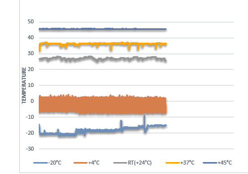
After drying at room temperature, each batch of Whatman paper was sealed in a zipper bag and incubated at different temperatures: +45°C, +37°C, +24°C, +4°C and -20°C respectively as shown in Figure 1. Incubation lasted successively 31 days, 24 days, 17 days, 10 days, and 3 days.
Polymerase chain reaction
At the end of the incubation period, all Whatman papers were collected and stored at -20°C before proceeding to nucleic acid extraction by batch.
A sample of Whatman paper was placed into an Eppendorf tube, followed by the addition of 500 µL of
RNase-free water. After vortexing, the tube was left in a Biosafety Cabinet (BSC) level 2 for one hour. Then, the solution was vortexed once more, and 140 µL was collected for RNA extraction using the QIAamp® Viral kit. (21).
During the extraction, the sample was first lysed with 560 µL of AVL buffer containing the carrier RNA in the 1.5 mL microcentrifuge tube, then fixed with ethanol (97%) 560µl in each tube. The resultant was washed with 500 µL of the wash buffer and AW2, and finally eluted with 60 µL of AVE buffer (21).
Five (05) µL of the eluate was then used to proceed to reverse transcript PCR using the Qiagen One Step PCR kit according to the manufacturer’s instructions (22). Primers NP3 and NP4 for detection of Morbillivirus RNA were used to amplify a conserved 351 bp region of the N gene (See Table 1). Then, the tubes were properly closed, labeled, and placed in the thermal cycler for RNA amplification according
to the program.
At the end of the program run, 5 µL of the amplified sample was mixed with 2 µL of loading buffer on paraffin paper before being transferred to the gel wells containing 1% agarose and 3% red gel. The migration was done for 1h with an intensity of 100 A (21).
Data analysis
The results were displayed in tabular form within an Excel spreadsheet.
The t-test of Stata software was used to analyze the variation in RNA titer readings and subsequently the mean was compared to the OIE titer standard. Additionally, Excel software was employed to assess the
temperature fluctuations during the incubation period.
Results
The different dilutions inoculated on the Whatman paper were incubated at various temperatures as shown in figure 1.
After 3 to 31 days of incubation, the samples of Whatman paper were collected and stored at -20°C. They were then subjected to RNA purification followed by RT-PCR amplification. The amplified RNA was then detected by electrophoresis on 1% agarose gel containing Gel-red and observed under UV.
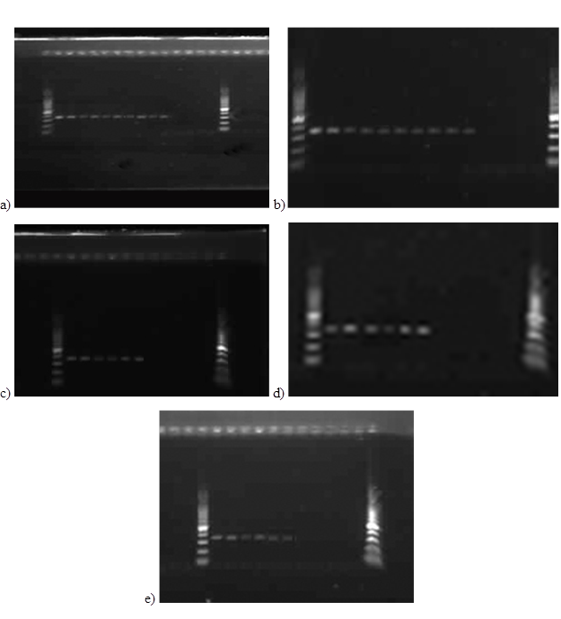
Serial dilutions were performed. Only the undiluted sample showed well-preserved RNA after 31 days storage. This was confirmed by the successful detection of PPR viral RNA (Tables 2 and 3). Figure 2 further demonstrates efficient amplification of the NP gene fragment from PPR virus isolates stored on Whatman paper at different temperatures: a) -20°C, b) +4°C, c) +24°C, d) +37°C, and e) +45°C. Amplification was performed using NP3/NP4 primers and visualized by electrophoresis on a 1% agarose gel.
The sample of dilution 0.1 with titer 101.46 TCID50/ml shows also well-preserved RNA after 31 day’s storage when incubate at -20°C and +4°C (Table 2) while at +24°C, +37°C and +45°C the RNA is only present during the first three days (Table 3).

Figure 1: Chart of temperature conditions
Table 1: Primer sequences of N-gene sequence of PPRV used for RT-PCR as described by Couacy-Hymann et al. (22)
| Primer ID | Purpose | Target | Location | Sequence (5’-3’) |
| NP3 | Forward | NP gene | 1232-1255 | TCT CGG AAA TCG CCT CAC AGA CTG |
| NP4 | Reverse | NP gene | 1585-1560 | CCT CCT CCT GGT CCT CCA GAA TCT |
Table 2: Thermo-stability of PPR Virus RNA at -20°C and +4°C by RT-PCR
| Vaccine dilutions | TCID50 | Duration of incubation | ||||
| Day 3 | Day 10 | Day 17 | Day 24 | Day 31 | ||
| 1 | 102.46 TCID50/ml | + | + | + | + | + |
| 0.1 | 101.46 TCID50/ml | + | + | + | + | + |
| 0.01 | 100.46 TCID50/ml | – | – | – | – | – |
| 0.001 | 10-1.46 TCID50/ml | – | – | – | – | – |
Table 3: Thermo-stability of PPR Virus RNA at +24°C, +37°C and +45°C by RT-PCR
| Vaccine dilutions | TCID50 | Duration of incubation | ||||
| Day 3 | Day 10 | Day 17 | Day 24 | Day 31 | ||
| 1 | 102.46 TCID50/ml | + | + | + | + | + |
| 0.1 | 101.46 TCID50/ml | + | – | – | – | – |
| 0.01 | 100.46 TCID50/ml | – | – | – | – | – |
| 0.001 | 10-1.46 TCID50/ml | – | – | – | – | – |

Figure 2: Specific PCR amplification of the NP gene fragment of different PPRV isolates from Whatman paper stored at a)-20°C, b)+4°C, c) +24°C d)+37°C, e)+45°C with primers NP3/NP4. The amplified products are analyzed by electrophoresis on 1% agarose gel stained with loading buffer and read under UV.
Discussion
This study focused on evaluating the potential of Whatman paper Grade 3MM CHR as a tool for preserving PPRV nucleic acid at various temperatures.
o get the PPR virus nucleic acid, we used the PPR vaccine. Similar experiments by other scientists demonstrated that whole blood (8) is the most convenient sample to collect on filter paper. However, many reference tests have used other types of samples (e.g., serum or plasma) (24), and some diseases are best diagnosed using other types of samples (7).
PCR analysis revealed that Whatman paper effectively preserved PPR virus nucleic acid at a 100 dilution throughout the incubation period. However, at a dilution 0.1, preservation was only successful at -20°C and +4°C for the entire experiment. At higher temperatures (+24°C, +37°C, and +45°C), the nucleic acid remained detectable only for the first three days (Figure 2). At even lower dilutions (0.01 and 0.001), RT-PCR amplification and agarose gel electrophoresis failed to detect any viral nucleic acid. This is likely due to the low concentration of infectious virus present in the Whatman paper at these dilutions (Table S1). Similar studies confirm this result. As example, in 2003, Bossuyt and collaborators demonstrated that hepatitis virus showed a 10 fold drop in viral load after storage for 4 weeks on Whatman paper at room temperature (25). Also, according to Yuguda and collaborators, the PPR vaccine maintained the viability
at room temperature for a sizable amount of time after being reconstituted in PBS. After more than 8 hours, the reconstituted vaccine’s titer of 103.1 TCID50 remained stable, but it began to decline after 15 hours at room temperature (26). In addition, according to a study performed by Kailash et al., in the framework of the human papillomavirus (HPV) study, the quality and quantity of DNA derived from paper
smears and the results of PCR amplifications for HPV type 16, BRCA1, and p53 genes from cervical smear samples, impression biopsies, blood and fine needle aspirations are stable in paper smears for up to one year stored at -70°C (12). This method is simple, rapid, and economical, and can be used effectively for large-scale population screening, especially in areas where samples must be transported from remote locations to the laboratory.
Conclusion
This research highlights the potential of Whatman paper Grade 3MM CHR as a tool for preserving PPRV nucleic acid, particularly at higher dilutions and lower temperatures. The study demonstrated that at a dilution of 1 (100-fold), Whatman paper effectively maintained viral nucleic acid integrity throughout the experimental period. However, at lower dilutions (0.1, 0.01, and 0.001), successful preservation was limited to colder temperatures (-20°C and +4°C) for dilution 0.1 only, with diminishing stability observed at higher temperatures. These findings underscore the importance of appropriate storage conditions and the sensitivity of preservation methods to dilution levels and environmental factors. Additionally, the study contributes to the body of knowledge regarding the stability of viral nucleic acids on filter paper substrates, offering insights valuable for disease surveillance, diagnostics, and vaccine storage strategies. Moving forward, further investigations into optimization strategies for preserving viral nucleic acids on
Whatman paper, as well as exploration of alternative storage methods, could enhance the utility and applicability of this preservation approach in various healthcare and research settings.
Abbreviations
3MM CHR : 0.34 millimeters “Chromatography”; AU PANVAC : African Union – Pan African Veterinary Vaccine Centre; AVL Buffer : A Viral Lysis Buffer ; BSC : Biosafety Cabinet ; DNA : Deoxyribonucleic acid ; EDTA : Ethylenediaminetetraacetic acid ; FAO : Food and Agriculture Organization of the United Nations ; FTA : Fast Technology for Analysis of nucleic acids ; GMEM : Glascow Minimum Essential Medium ; IATA : International Air Transport Association ; OIE : Office International des Épizooties ; PBS : Phosphate Buffered Saline ; PCR : Polymerase Chain Reaction ; PPR : Peste des Petits Ruminants ; PPRV : Peste des Petits Ruminants Virus ; RNA : Ribonucleic acid ; RT-PCR : Reverse-Transcription Polymerase Chain Reaction ; TCID50 : Median Tissue Culture Infectious Dose ; UV : Ultraviolet
Acknowledgements
We appreciate Mrs. Rahamatou CISSE of AU-PANVAC for stimulating orderliness towards the success of this project. We also highly indebted to the staff of African Union Pan African Veterinary Vaccine Centre (AU PANVAC), Debre Zeit, Ethiopia staff, and express our deep gratitude to Professor Gabriel OGUNDIPE for the supervision.
Funding
This work was fully funded by the African Union and the
Pan-African University.
Conflict of interest
There is no conflict of interest.
Author’s contribution
Akakpo Oluwole Christophert: Designed the study methodology, performed most of data collection and analysis and drafted the manuscript.
Bodjo Sanne Charles (DVM): Oversaw drafting of study protocol, analysed the data, and led overall research project in the laboratory at AU-PANVAC in Ethiopia
Diallo Safiatou Lamine (DVM, MSc): Edited the manuscript before submission.
References
- WOAH. Peste des petits ruminants – WOAH – World Organisation for Animal Health [Internet]. 2018 [cited Aug 8, 2022]. Available on: https://www.woah.org/en/disease/peste-des-petits-ruminants/
- OIE. Peste des petits ruminants – OIE – World Organisation for Animal Health. 2021
- Adombi CM, Lelenta M, Lamien CE, Shamaki D, Koffi YM, Traoré A, et al. Monkey CV1 cell line expressing the sheep–goat SLAM protein: a highly sensitive cell line for the isolation of peste des petits ruminants virus from pathological specimens. Journal of virological methods.
2011;173(2):306-13 - Balamurugan V, Hemadri D, Gajendragad M, Singh R, Rahman H. Diagnosis and control of peste des petits ruminants: a 2014;25:39-56 comprehensive review. Virus disease.
- Kamel M, El-Sayed A. Toward peste des petits virus (PPRV) eradication: Diagnostic approaches, novel vaccines, and control strategies. Virus research. 2019;274:197774
- Hossain MT, Rahman M, Begum J, Haque M, Engineering. Development and Validation of Filter Paper
Method of RT-PCR for the Detection of ‘N’gene of Peste Des Petits Ruminants Virus. American Journal of Biomedical Science. 2017;3(4):37-44 - Li C-C, Beck IA, Seidel KD, Frenkel LM. Persistence of human immunodeficiency virus type 1 subtype B DNA in dried-blood samples on FTA filter paper. Journal of clinical microbiology. 2004;42(8):3847-9
8.Chaisomchit S, Wichajarn R, Janejai N, Chareonsiriwatana W. Stability of genomic DNA in dried blood spots stored on filter paper. Southeast Asian J Trop Med Public Health. 2005;36(1):270-3 - Prado I, Rosario D, Bernardo L, Álvarez M, Rodríguez R, Vázquez S, et al. PCR detection of dengue virus using dried whole blood spotted on filter paper. Journal of virological methods. 2005;125(1):75-81
- Yourno J, Conroy J. A novel polymerase chain reaction method for detection of human immunodeficiency virus in dried blood spots on filter paper. Journal of clinical
microbiology. 1992;30(11):2887-92 - Pitcovski J, Shmueli E, Krispel S, Levi N. Storage of viruses on filter paper for genetic analysis. ournal of virological methods. 1999;83(1-2):21-6 Kailash U, Hedau S, Gopalkrishna V, Katiyar S, Das B. A simple ‘paper smear’method for dry collection, transport and storage of cervical cytological specimens for rapid screening of HPV infection by PCR. Journal of medical microbiology. 2002;51(7):606-83
- Nwankpa N, Bodjo C, Tounkara K, Domenech J. Quality control of PPR vaccine in Africa: the role of AU-PANVAC. OIE Bull. 2015;2:64-71
- Word Health Organization. Global plan to combat neglected tropical diseases 2008-2015. World Health Organization; 2007
- Bildfell R. Overview of Collection and Submission of Laboratory Samples. Accessed 15th June 2019
- Organization WH. Guidance on regulations for the transport of infectious substances 2021-2022: applicable as from 1 January 2021. 2021
- Leppard A. NHS Foundation Trust. 2013 [cited Aug 10, 2022]. Transport of Specimens to the Laboratory-Pathology. Available on : https://www.barnsleyhospital.nhs.uk/pathology/pathology
general/transport-spceimens-laboratory/ - Bhuiyan AR, Chowdhury EH, Kwiatek O, Parvin R, Rahman MM, Islam MR, et al. Dried fluid spots for peste des petits ruminants virus load evaluation allowing for non invasive diagnosis and genotyping. BMC Veterinary Research. 2014;10:1-8
- Philip B, Brian B. Ethiopia [Internet]. Bradt Travel Guides; 2009 [cited Aug 3, 2022]. p. 614. Available on: https://books.google.com/books/about/Ethiopia.html?id=QFb 6pacaczsC
- Martrenchar A, Zoyem N, Diallo A. Experimental study of a mixed vaccine against peste des petits ruminants and capripox infection in goats in northern Cameroon. Small Ruminant Research. 1997;26(1-2):39-44
- Chukwudi IC, Ogbu KI, Luka PD, Malesa RP, Heath LE, Ugochukwu EI, et al. Comparison of colorimetric loop mediated isothermal amplification kit and reverse transcription-polymerase chain reaction in the diagnosis of peste des petits ruminants in sheep and goats in Southeast Nigeria. Veterinary World. 2020;13(11):2358
- Couacy-Hymann E, Roger F, Hurard C, Guillou J, Libeau G, Diallo A. Rapid and sensitive detection of peste des petits ruminants virus by a polymerase chain reaction assay. Journal of virological methods. 2002;100(1-2):17-25
- Julho. Tissue Culture Infectious Dose (TCID50) Assays | BMG LABTECH. LABTECH. 2021;7
- Kalayou S, Tadelle H, Bsrat A, Abebe N, Haileselassie M, Schallig H. Serological evidence of Leishmania donovani infection in apparently healthy dogs using direct agglutination test (DAT) and rk39 dipstick tests in Kafta Humera, north‐west Ethiopia. Transboundary emerging
diseases. 2011;58(3):255-62 - Bossuyt PM, Reitsma JB, Bruns DE, Gatsonis CA, Glasziou PP, Irwig LM, et al. Towards complete and
accurate reporting of studies of diagnostic accuracy: the STARD initiative. 2003 - El-Yuguda A-D, Baba SS, Ambali AG, Egwu GO. Field trial of a thermostable peste des petits ruminants (PPR) vaccine in a semi-arid zone of Nigeria. World Journal of
Vaccines. 2014;04(01):1–6
